India ink staining, also known as negative staining, is a simple yet effective approach for visualizing the exterior structures of bacteria, fungi, and other microbes in microbiology. The dye is used to stain the background in this staining process, while the microbe shows as a clear patch against the dark background. We will explore the principle, technique, benefits, restrictions, comparison with other stains, and quality control of India ink staining in this article.
Cryptococcal meningitis occurs in immunocompromised patients and when meningitis is clinically suspected, such as in HIV patients, or when yeast cells with lymphocytes are detected when performing a C.S.F. cell count or examining a Gram smear, or when India ink preparation for encapsulated yeasts is examined.
India ink is used as a negative stain in ion-negative staining, which allows visualization of the normally transparent and unstainable capsules of various microorganisms such as Cryptococcus neoformans (most commonly), Klebsiella pneumoniae, Streptococcus pneumoniae, and others.
-
Questioned Document MCQs Part 2 | E...Loaded: 95.43%Remaining Time 1:34
Principle
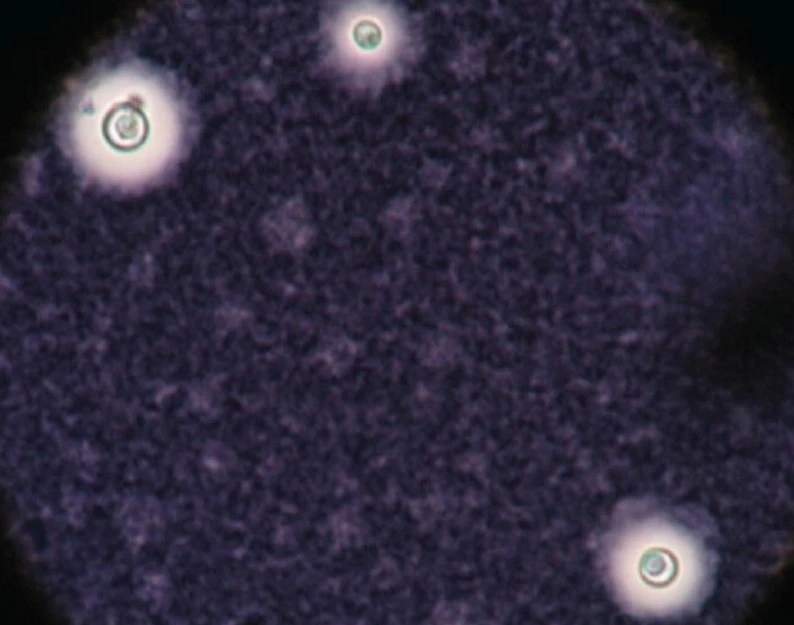
The India ink staining technique is based on the negative staining principle, which states that the dye is used to stain the background while the microbe remains unstained, resulting in a clear image against a dark background. The India ink stain is made up of microscopic, opaque carbon black particles floating in an aqueous solution. Because the carbon black particles do not penetrate the microbe, it remains unstained and visible under the microscope. Because the capsule is non-ionic, the India ink used will not bind to it. As a result, the capsule appears as a clear halo around the yeast cells.
India Ink Composition
Deionized Water, Thimerosal and Black Pelican Drawing Ink No. 17
Requirements for India Ink Preparation
- Nigrosine stain or India ink,
- Slides and coverslips that are clean and free of grease.
- The C.S.F. specimen,
- Droppers or a loop for inoculation,
- Bunsen burner
- waste disposal container,
- Centrifuge,
- Tubes for testing,
- Microscope and
- Cryptococcus neoformans (for positive control)
- Candida albicans used for negative control.
Ink preparation
- CSF should be centrifuged for 5 to 10 minutes.
- Remove the supernatant fluid and combine it with the sediment.
- Transfer an equal amount of sediment and India ink to a slide, i.e. a drop of sediment and a drop of India ink.
- Cover with a coverslip after mixing. Examine the preparation using the 40 X objective under a microscope.
Observation
Look for oval or round cells that are irregular in size, measuring 2-10 m in diameter, and are surrounded by a large unstained capsule.
Result interpretations
Positive control: the presence of encapsulated yeasts was observed.
Negative control: The absence of encapsulated yeasts was observed.
Advantages
There are various advantages of using India ink staining, including:
- it can be performed easily.
- Minimal equipment and materials are required.
- A vivid, high-contrast view of the microorganism's exterior structures is provided.
- It is capable of displaying a vast variety of microorganisms, including bacteria, fungus, and parasites.
- Heat fixation is not required, which might harm fragile structures.


